
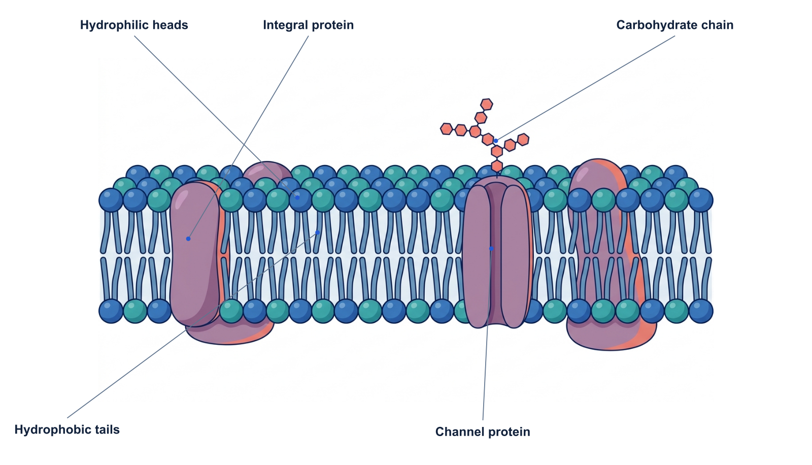
A simplified AI-generated membrane cross section. Colors separate components; the membrane is dynamic, and the picture does not show every molecule present.
The plasma membrane is a selective boundary built around a phospholipid bilayer. Its structure allows a cell to maintain an internal environment while communicating and exchanging materials with its surroundings.
In this cross section, water-facing heads sit on the outer surfaces of the bilayer. The hydrophobic tails face inward. Proteins and attached carbohydrates add functions that the lipid barrier alone cannot provide.
Parts and functions
| Structure | What it does |
|---|---|
| Hydrophilic heads | Water-interacting portions of phospholipids facing the aqueous environments. |
| Hydrophobic tails | Nonpolar fatty-acid regions oriented toward the bilayer’s interior. |
| Integral protein | A protein embedded in the membrane; many span the bilayer. |
| Channel protein | A membrane protein that provides a route for specific substances to cross. |
| Carbohydrate chain | An extracellular chain attached to a protein or lipid, involved in functions such as recognition. |
Why phospholipids form a bilayer
A phospholipid has regions with different interactions with water. In the cell’s aqueous environment, this arrangement favors a structure that exposes the heads to water and shelters the tails inside.
The bilayer is not a rigid wall. Many components can move laterally, and membrane properties depend on its composition and conditions. That combination of mobility and mixed components is reflected in the fluid-mosaic model.
Selective permeability is more than a hole in a wall
The lipid interior presents a barrier to many charged and polar substances. Channels, carriers and other proteins provide controlled ways to move substances across it. A channel is not simply an empty gap between phospholipids.
Different membrane proteins have different roles: transport, signaling, attachment and enzymatic activity are examples. A single generic protein shape does not identify a specific receptor or channel.
Teach the two sides clearly
The carbohydrate chain is shown on the extracellular side. If you rotate or crop the illustration, preserve that distinction and label the relevant environments explicitly.
For a teaching sequence, introduce the bilayer first, then the proteins, then recognition or transport. Use the unlabeled view to ask which part of a phospholipid faces water.
A prompt to start from
Frequently asked questions
References and further reading
Illustrations are explanatory models. Colors, proportions and selected structures are simplified to support the topic.
Keep exploring
Try it free
Start with a figure. Make it yours.
Open a starter, adapt the labels, and create an illustration for your next lecture or research discussion.
Starter examples are free to open. AI generation and edits use credits.