
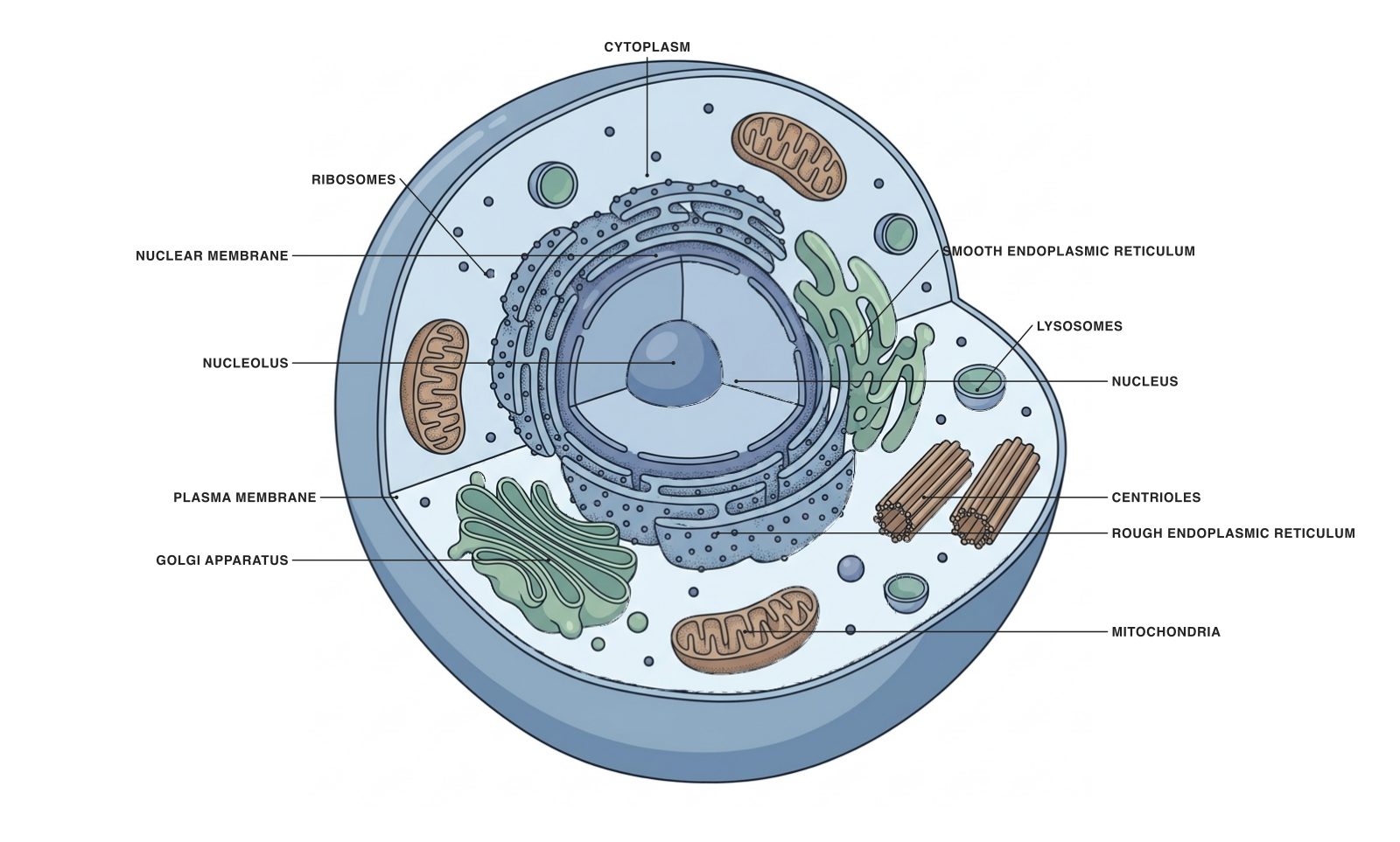
A generalized animal cell created in Underleaf Figure Maker. Structures are selected and enlarged for explanation; actual cells vary by tissue.
An animal cell has a plasma membrane, a nucleus and specialized internal compartments. This diagram shows a generalized cell rather than a particular cell type, making it useful for introducing the organelles shared by many animal cells.
Read the labels as a map of different jobs. Some structures manage genetic information, some make or process proteins, and others support metabolism, transport or organization.
Parts and functions
| Structure | What it does |
|---|---|
| Plasma membrane | A selective boundary that separates the cell from its environment. |
| Nucleus | Houses most of the cell’s genetic material. |
| Nuclear envelope | A double membrane surrounding the nucleus; nuclear pores regulate exchange. |
| Nucleolus | Helps assemble ribosomal subunits. |
| Ribosomes | Synthesize proteins using information carried by messenger RNA. |
| Rough ER | Produces and begins processing many proteins destined for membranes or secretion. |
| Smooth ER | Supports lipid synthesis and, in some cells, calcium storage or detoxification. |
| Golgi apparatus | Modifies and sorts products for delivery to cellular destinations. |
| Mitochondria | Generate much of the ATP used by aerobic cells. |
| Lysosomes | Digest and recycle material using enzymes in an acidic compartment. |
| Centrioles | Components associated with the centrosome in many animal cells. |
| Cytoplasm | The cellular region outside the nucleus, containing cytosol and organelles. |
Read the diagram as a protein-making route
A useful teaching sequence is nucleus → ribosome → rough ER → Golgi apparatus. It connects several labels with one process instead of presenting the organelles as an isolated vocabulary list.
This route applies to many secreted and membrane proteins. It is not the route for every protein: free ribosomes make many proteins that work in the cytosol or are directed elsewhere. Do not draw a single universal path for all protein synthesis.
What an animal cell does not show
This animal-cell model does not have a cellulose cell wall, chloroplasts or a large plant-like central vacuole. It still has a plasma membrane, and it still uses compartmentalization to organize chemistry.
A real neuron, muscle cell or red blood cell will look different from this general-purpose diagram. For example, mature mammalian red blood cells lack a nucleus. Use the generalized diagram to establish a baseline before introducing specialized cells.
Adapt the labeled and unlabeled versions
Use the labeled diagram when introducing organelles. Use the unlabeled download for retrieval practice: ask students to place names, then explain a function without looking back at the table.
For a slide, reduce the labels to the structures discussed in that moment. A set of consistent diagrams with six labels per view is often easier to teach from than one image carrying every detail.
- Locate the nucleus and explain the nuclear envelope.
- Distinguish the rough ER from the smooth ER.
- Connect lysosomes with digestion and recycling rather than ATP production.
A prompt to start from
Frequently asked questions
References and further reading
Illustrations are explanatory models. Colors, proportions and selected structures are simplified to support the topic.
Keep exploring
Try it free
Start with a figure. Make it yours.
Open a starter, adapt the labels, and create an illustration for your next lecture or research discussion.
Starter examples are free to open. AI generation and edits use credits.