
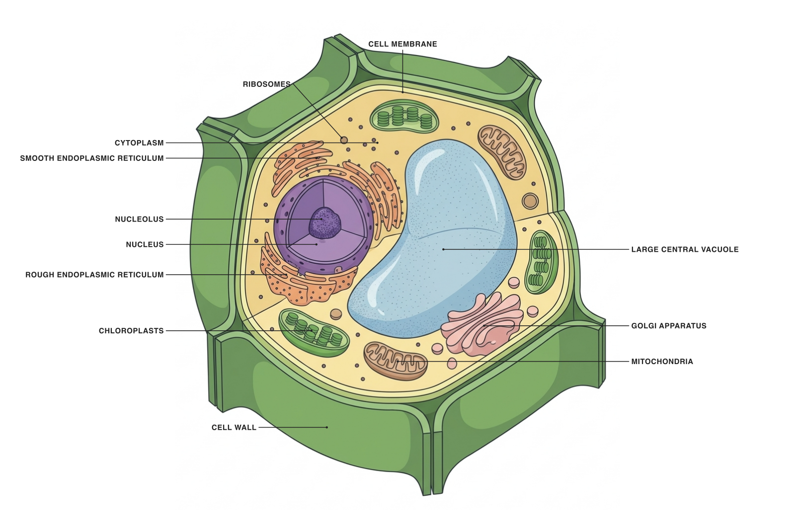
A generalized plant cell illustration from Underleaf Figure Maker. This is a conceptual cutaway, not a microscope image or a scale drawing.
A plant cell is a eukaryotic cell: its DNA is enclosed in a nucleus, and internal membranes organize many of its activities. This diagram brings the major structures into one view so you can connect each label with its role.
The cell wall, chloroplasts and large central vacuole make a useful starting point for comparison with an animal cell. The illustration represents a photosynthetic plant cell; cells from roots and other tissues do not all have the same collection of visible structures.
Parts and functions
| Structure | What it does |
|---|---|
| Cell wall | A supporting layer outside the plasma membrane. In plants, cellulose is a major structural component. |
| Cell membrane | The selectively permeable boundary controlling exchange between the cell and its surroundings. |
| Central vacuole | A large fluid-filled compartment involved in storage and turgor pressure. |
| Nucleus | Contains most of the cell’s DNA and supports the regulation of gene expression. |
| Nucleolus | A region within the nucleus involved in making ribosomal subunits. |
| Chloroplasts | Organelles in which photosynthesis converts light energy into chemical energy. |
| Mitochondria | Organelles that support cellular respiration and ATP production. |
| Ribosomes | Structures that translate messenger RNA into proteins. |
| Rough endoplasmic reticulum | A membrane network associated with ribosomes and the production of many secreted and membrane proteins. |
| Smooth endoplasmic reticulum | A membrane network involved in lipid synthesis and other functions that vary by cell type. |
| Golgi apparatus | Modifies, sorts and packages proteins and lipids. |
| Cytoplasm | The material inside the plasma membrane and outside the nucleus, including cytosol and organelles. |
How to recognize the main structures
Start at the edge. The cell wall sits outside the thin plasma membrane; the two labels refer to different structures. Move inward to the large central vacuole, then locate the nucleus and smaller organelles around it.
Look for chloroplasts and mitochondria separately. Plant cells need both photosynthesis and cellular respiration; a chloroplast does not replace a mitochondrion. The colors here distinguish structures and should not be interpreted as their natural appearance.
Plant cells and animal cells
Both have a nucleus, a plasma membrane, mitochondria, ribosomes, endoplasmic reticulum and a Golgi apparatus. A typical animal-cell diagram lacks the plant cell wall, chloroplasts and large central vacuole.
Shape alone is a weak test. Real plant and animal cells vary with their tissue and function, while textbook diagrams deliberately emphasize recognizable features.
Use the unlabeled diagram as a worksheet
Switch to the unlabeled view and download it for a class activity. Ask learners to identify the boundary, genetic compartment and energy-related organelles before adding every label. Use the labeled view and parts table as the answer key.
For an introductory lecture, use fewer labels and explain one relationship at a time. For a more detailed session, expand the discussion to membrane trafficking, compartmentalization or the link between chloroplasts and photosynthesis.
- Identify the structure that maintains turgor pressure.
- Explain why a plant cell contains mitochondria as well as chloroplasts.
- Trace the outside-to-inside order of cell wall, membrane and cytoplasm.
A prompt to start from
Frequently asked questions
References and further reading
Illustrations are explanatory models. Colors, proportions and selected structures are simplified to support the topic.
Keep exploring
Try it free
Start with a figure. Make it yours.
Open a starter, adapt the labels, and create an illustration for your next lecture or research discussion.
Starter examples are free to open. AI generation and edits use credits.