
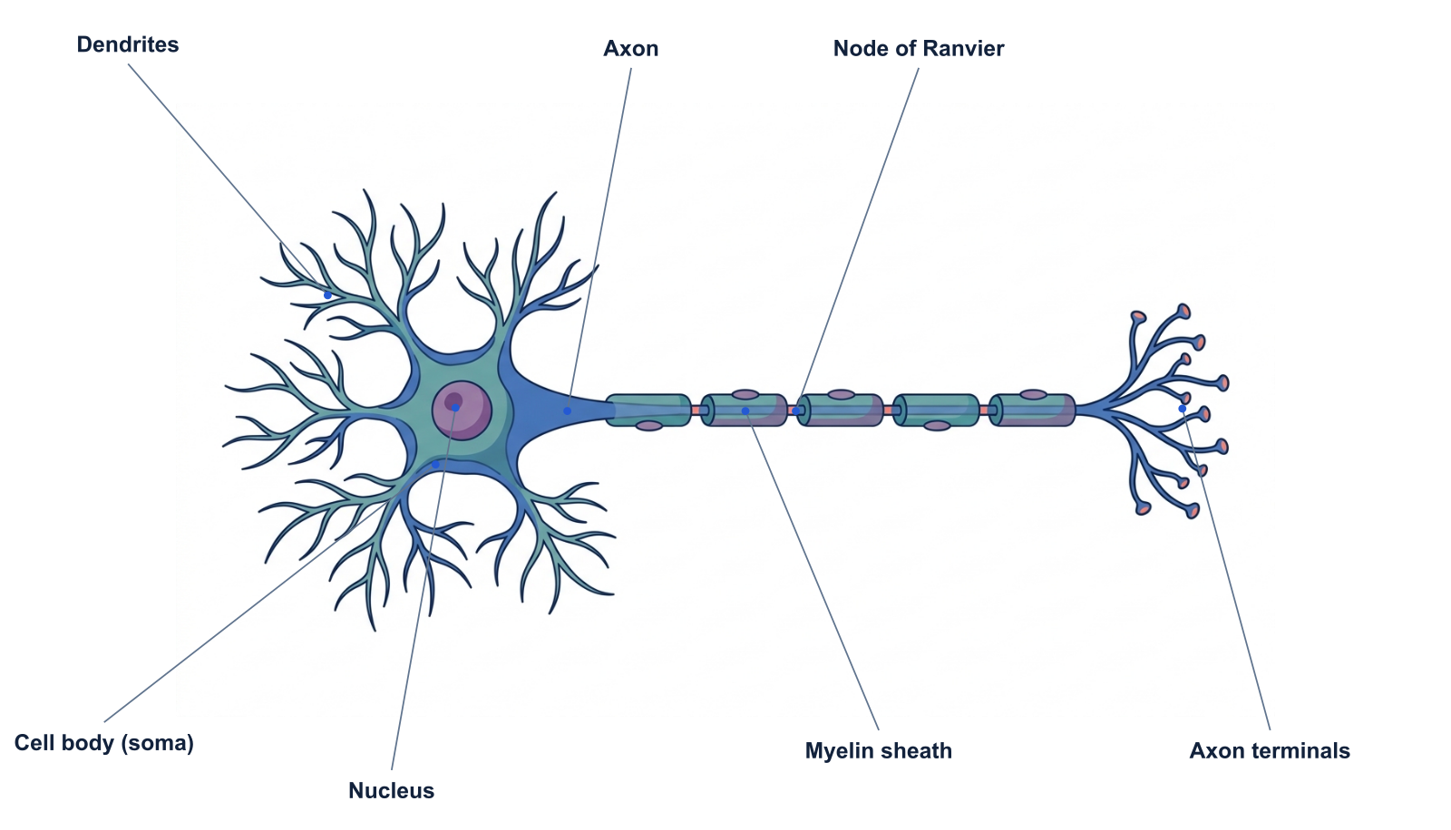
AI-generated teaching illustration with separately placed labels. This schematic represents a myelinated multipolar neuron; it is not a universal shape for all neurons.
A neuron is a specialized cell that receives, integrates and transmits information. The long shape in this diagram emphasizes how different regions contribute to communication.
The illustration shows a common introductory model: many dendrites near a cell body, one long myelinated axon, and branches ending in terminals. Neurons in different circuits can have very different shapes.
Parts and functions
| Structure | What it does |
|---|---|
| Dendrites | Receive many of the inputs arriving from other cells. |
| Cell body (soma) | Contains the nucleus and supports much of the cell’s metabolism. |
| Nucleus | Contains genetic material and supports regulation of cellular activity. |
| Axon | Conducts action potentials toward distant targets. |
| Myelin sheath | An insulating covering made by glial cells around segments of many axons. |
| Node of Ranvier | A gap between myelin segments where action potentials are regenerated during saltatory conduction. |
| Axon terminals | Specialized endings involved in communicating with a target cell. |
Follow the general direction of communication
For an introductory chemical-synapse model, follow information from dendritic inputs toward the cell body and along the axon to its terminals. The action potential is normally initiated near the beginning of the axon after inputs have been integrated.
This is a teaching model, not a rule describing every signal in every neuron. If your topic is a specific circuit or cell class, label the actual connection and signaling direction rather than assuming every branch is equivalent.
Why myelin and nodes belong together
Myelin wraps segments of an axon. The gaps between segments are the nodes of Ranvier. Treating the entire axon as one uninterrupted sheath hides the relationship between these two structures.
In a lecture about conduction, first show the axon and sheath. Then emphasize the nodes. Do not infer conduction velocity from the spacing in this picture; its lengths and diameters are not to scale.
Make an anatomy slide and a signaling slide
Use the labeled version to introduce the structures. Duplicate the explanation for a second slide with fewer labels and a single clear indication of signal direction. Keeping the same geometry helps the audience focus on the new concept.
For a worksheet, download the unlabeled view and ask learners to name the gaps in the sheath as well as the sheath itself. The distinction is a useful check of understanding.
A prompt to start from
Frequently asked questions
References and further reading
Illustrations are explanatory models. Colors, proportions and selected structures are simplified to support the topic.
Keep exploring
Try it free
Start with a figure. Make it yours.
Open a starter, adapt the labels, and create an illustration for your next lecture or research discussion.
Starter examples are free to open. AI generation and edits use credits.