
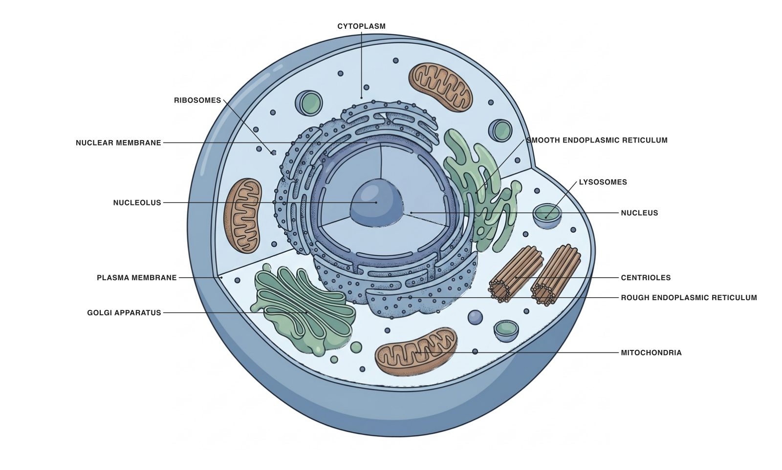
This animal cell illustrates eukaryotic organization. It is one example, not a diagram of every plant, fungal or protist cell.
A eukaryotic cell organizes its genetic material in a membrane-bound nucleus and divides many internal activities among specialized compartments. Animals, plants, fungi and diverse protists are eukaryotes.
The animal-cell illustration is a useful first example, but eukaryotic does not mean animal. The plant-cell model is equally eukaryotic, and it adds structures associated with plant biology.
Parts and functions
| Structure | What it does |
|---|---|
| Nucleus and nuclear envelope | A DNA-containing compartment bounded by a double membrane. |
| Endoplasmic reticulum | A membrane system involved in producing proteins and lipids. |
| Golgi apparatus | Processes and sorts cellular products. |
| Mitochondria | Support energy metabolism in the general eukaryotic model. |
| Plasma membrane | Separates the cell from its external environment. |
| Ribosomes | Produce proteins; ribosomes also occur in prokaryotic cells. |
Why compartmentalization matters
Membranes let different processes occur in different local environments. A lysosome, for instance, maintains conditions suited to its digestive functions, while the nucleus organizes processes involving genetic information.
When teaching this concept, connect each compartment with a specific activity. A labeled image becomes more meaningful when students can explain why the boundaries matter, rather than simply recall the names.
Animal, plant and other eukaryotic cells
Plant and animal cells share a nucleus, ribosomes and many membrane-bound organelles. Plant-cell diagrams usually emphasize the cell wall, chloroplasts and central vacuole; those features are not required for a cell to be eukaryotic.
Fungi and protists add further diversity. Avoid using a single diagram as a checklist that every mature cell must satisfy visibly: specialized cells can lose structures or modify them substantially.
The prokaryotic comparison
A prokaryotic cell has DNA, ribosomes and a plasma membrane, but no membrane-bound nucleus. The absence of a nucleus does not mean the absence of genetic organization.
For a side-by-side teaching figure, highlight the nuclear envelope first. Then compare the arrangement of internal membranes and the organelles relevant to the lesson. Keep image scales explicit if you introduce size comparisons.
A prompt to start from
Frequently asked questions
References and further reading
Illustrations are explanatory models. Colors, proportions and selected structures are simplified to support the topic.
Keep exploring
Try it free
Start with a figure. Make it yours.
Open a starter, adapt the labels, and create an illustration for your next lecture or research discussion.
Starter examples are free to open. AI generation and edits use credits.