
Biology / Cell structure
Cell diagrams, side by side.
Start with the shared functions. Then compare the structures that distinguish a generalized plant cell, animal cell and bacterial cell.
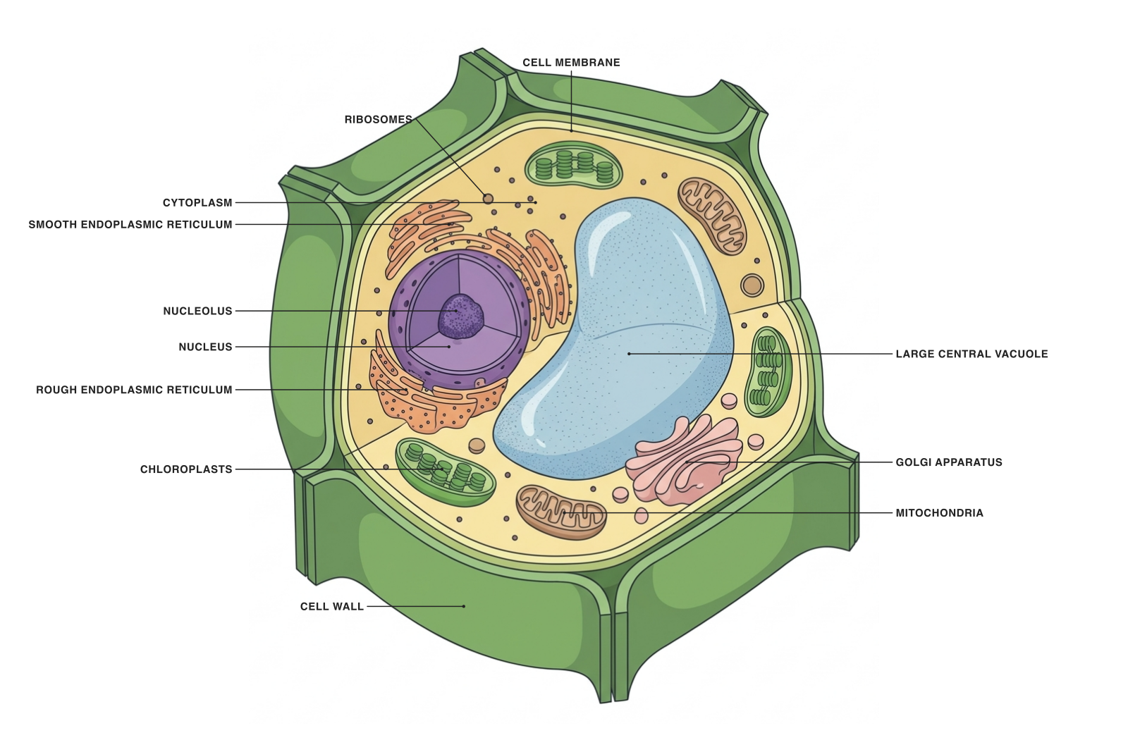
Plant cell
Cell wall, central vacuole and chloroplasts in a photosynthetic plant-cell model.
Explore
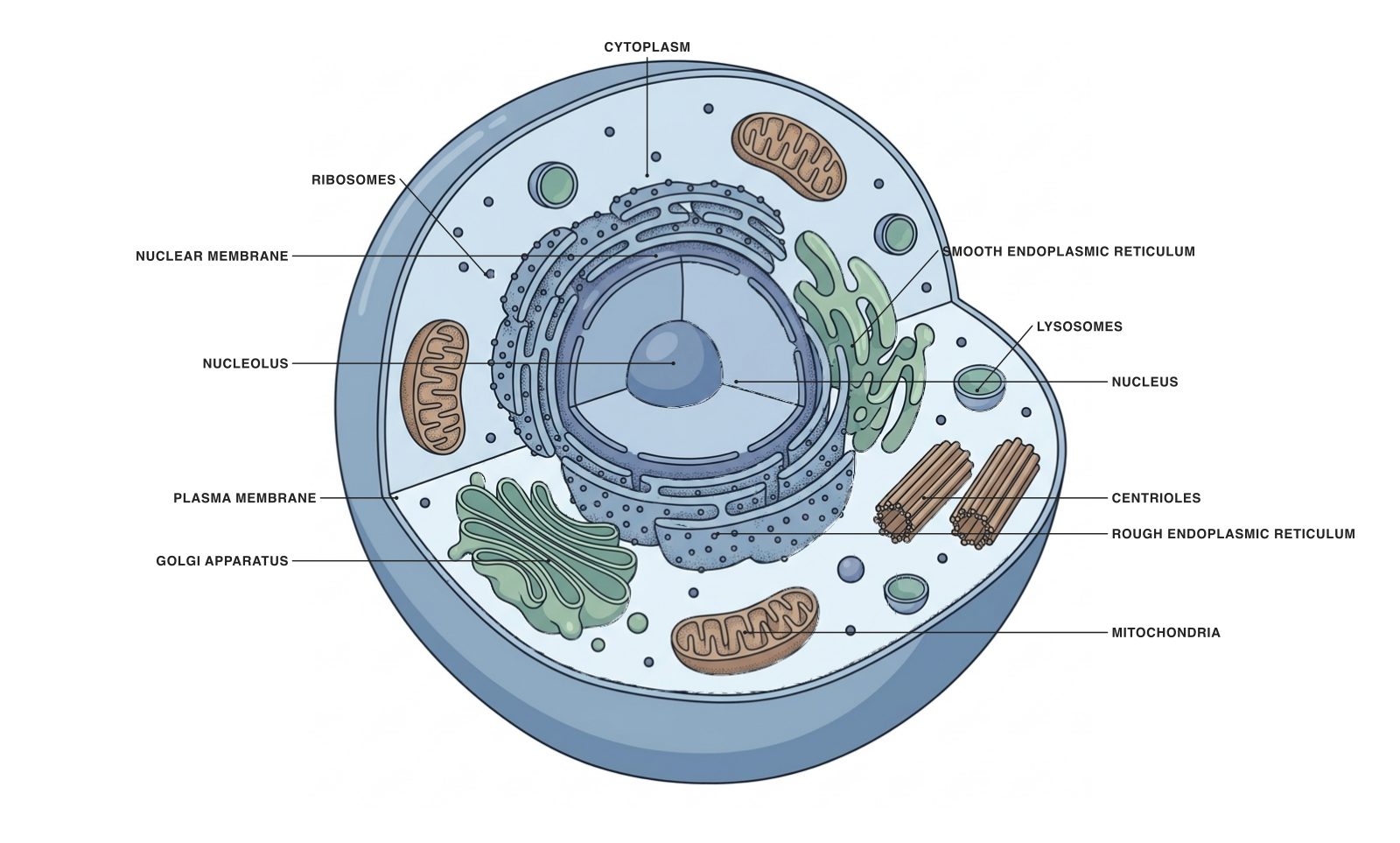
Animal cell
The nucleus and organelles of a generalized animal cell.
Explore
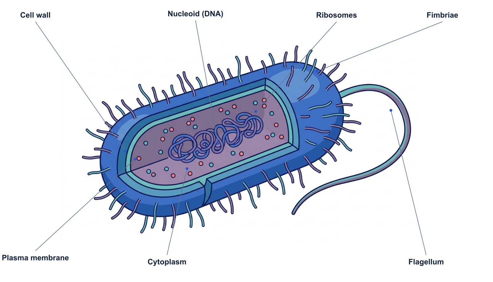
Prokaryotic cell
A bacterial model with a nucleoid and no membrane-bound nucleus.
Explore
What these cell diagrams have in common
Each cell needs a boundary, genetic information, a means of making proteins and an internal environment where reactions can occur. The diagrams therefore share a plasma membrane, DNA, ribosomes and cytoplasm, even though the organization differs.
The figures are explanatory models at different illustrative scales. Do not compare their drawn widths to infer real cell sizes. They also combine selected structures rather than representing every cell in a group.
Plant, animal and prokaryotic cells compared
| Feature | Plant model | Animal model | Bacterial model |
|---|---|---|---|
| Plasma membrane | Yes | Yes | Yes |
| Ribosomes | Yes | Yes | Yes |
| Membrane-bound nucleus | Yes | Yes | No |
| Mitochondria | Yes | Yes | No |
| Chloroplasts | In photosynthetic cells | No | No chloroplasts |
| Cell wall | Usually; cellulose-rich | No | Present in many; different composition |
| Large central vacuole | Typical of mature plant cells | No comparable central vacuole | No |
| Main DNA-containing region | Nucleus | Nucleus | Nucleoid |
The table describes general models. Specialized cells and different organisms introduce exceptions. For example, a root cell need not contain chloroplasts, and a mature mammalian red blood cell lacks a nucleus.
Eukaryotic versus prokaryotic is a different comparison
Plant and animal cells are both eukaryotic. The defining distinction in this introduction is the membrane-bound nucleus, together with the broader organization of internal compartments.
A cell wall does not decide the category: plants and many prokaryotes have walls, while animal cells do not. Likewise, the absence of a nucleus does not mean a prokaryote has no DNA.
Choose a useful diagram for your class
For a first comparison, use a short list: membrane, nucleus or nucleoid, ribosomes and the structures characteristic of the chosen model. Add organelles when the lesson requires their functions.
The individual diagram pages include labeled and unlabeled downloads and parts tables. For a worksheet, use the unlabeled views and ask students to explain one similarity and one difference, rather than only matching names to arrows.
References: OpenStax prokaryotic cells and eukaryotic cells.
Try it free
Adapt a cell diagram to your explanation.
Start with the plant or animal cell, change the labels, and build a version suited to the lesson you are teaching.
Starter examples are free to open. AI generation and edits use credits.